Stop Squinting to See
Schedule An Eye Exam In Lafayette, LA
Do you have frequent headaches? Is your vision blurry? Have you been rubbing your eyes frequently? If you answered yes to any of those questions, you need an eye exam. All of these symptoms and many more can be caused by vision changes. The trained optometrists at Today’s Eyecare will check your eyes for any health or vision problems.
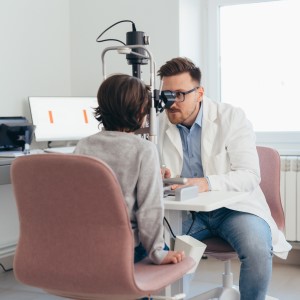
During your comprehensive eye exam we will test your eye movements, determine if a prescription is needed for glasses or contacts as well as assess the health of your eye. To make sure you’re seeing clearly, schedule an eye exam today by calling (337) 313-2020.
Looking for an eye doctor in Lafayette, LA? We’ve been in Lafeyette for 30 years but have just moved to a new state-of-the-art clinic.

Are your eyes healthy?
If you don’t know the answer, make an appointment with an eye care professional at Today’s Eyecare. Besides eye exams, residents of the Lafayette, LA area come to us for medical testing. We offer state-of-the-art testing for treatment and diagnoses of:
There’s more to your eye care/health than just vision

When you have an eye emergency in Lafayette, LA
Today’s Eyecare is open for eye emergencies, even on Saturdays. We can help with things like:
As soon as you notice a problem, don’t wait to call or walk in today (337) 313-2020. We are centrally located in Lafayette near Acadina Mall, close to Johnston and Ambassador.
GET TREATED QUICKLY
You use your eyes every day. An issue with your eyes can throw the whole day off track. Many residents of Lafayette, LA go to Today’s Eyecare because we’re open on the weekends. You can visit our emergency eye care center at a time that’s convenient for you.
Call us at 337-313-2020 right now to make an appointment.

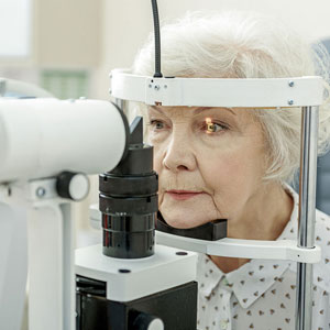
Slit Lamp Test
Allows your doctor a highly magnified view of your eye to thoroughly evaluate the front structures of your eye (lids, cornea, iris, etc.), followed by an examination of the inside of your eye (retina, optic nerve, macula and more). This test aids the doctor in the diagnosis of cataracts, dry eyes, corneal irritation, glaucoma and age-related macular degeneration.
Optomap Ultra-widefield Retinal Imaging
Creates a digital image that captures more than 80% of your retina in one panoramic image. Helps detect early signs of retinal disease including age-related macular degeneration, glaucoma and diabetic retinopathy.
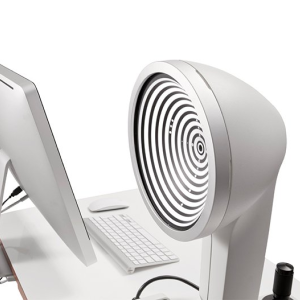
Oculus Keratograph 5M
The new Keratograph® 5M technology is a revolution in precision corneal topography and dry eye analysis. The high-resolution color camera and the integrated magnification changer offer a new perspective to the tear film assessment procedure for dry eye analysis. These detailed maps allow our doctors to precisely fit specialty contacts for irregular corneas and more.
Visual Fields Test
Checks for the presence of blind spots in your peripheral, or “side”, vision. These types of blind spots can originate from eye diseases such as glaucoma. Analysis of blind spots also may help identify specific areas of brain damage caused by a stroke or tumor. These tests are vital in assessing visual function for all types of eye conditions.
Color Vision Test
Evaluates color deficiencies in the eyes (red/green or blue/yellow) by asking you to pick out numbers from colored mosaic-like illustrations. In addition to detecting hereditary color vision deficiencies, the results may also alert your doctor to possible eye-health problems that could affect your color vision.
Specular Microscope
A camera that takes high-magnification images of the cellular layer of the inner surface of the cornea. It is used, to help evaluate the healing of the corneal epithelium after corneal injury, specific diseases or corneal surgery such as a keratectomy.
Ocular Coherence Tomography (OCT)
Takes cross-sectional pictures of the retina via a scanning laser. This technology is used to help diagnose and follow treatment in certain eye conditions and diseases, such as age-related macular degeneration, glaucoma and diabetic retinopathy.